Cases Study

▋ Introduction
Any clinician with implant experience knows that when a patient presents with very loose (low-density) alveolar bone, it can be a real nightmare. Not only does the implant fail to achieve sufficient primary stability after placement, but more concerning is that the implant may “float” within the soft bone. In severe cases, it can even migrate into the sublingual space or drop into the maxillary sinus, essentially becoming lost.
Oral and maxillofacial surgeons frequently encounter such cases referred to emergency departments or hospitals. Today, I will not focus on how to manage these challenging complications. Instead, I would like to discuss how to predict poor bone quality in advance so that adequate preparation can be made early on.
▋ Importance of Imaging Interpretation
First and foremost, proper interpretation of imaging is critical. On CBCT grayscale images, areas with a higher proportion of dark regions—or larger dark zones—should raise concern. While this may sometimes be influenced by imaging dose, a more careful evaluation often reveals underlying issues.
In addition, imaging software should be utilized effectively to aid assessment. One useful parameter is the CT number, also known as the Hounsfield Unit (HU) value. This objective measurement provides valuable information, although relatively few clinicians routinely evaluate it.
In 1999, Carl Misch proposed a classification based on HU values:
D1 bone: >1250 HU
D2 bone: 850–1250 HU
D3 bone: 350–850 HU
D4 bone: 150–350 HU
D5 bone: <150 HU
▋ Examples
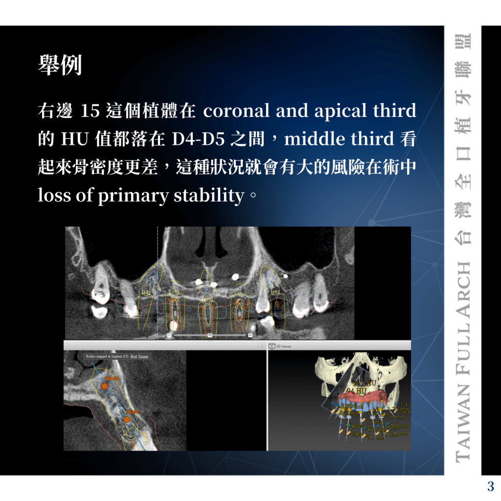
In Figure 1 (right side, implant #15), the HU values at both the coronal and apical thirds fall within D4–D5. The middle third appears to have even lower bone density. In such cases, there is a significant risk of intraoperative loss of primary stability.
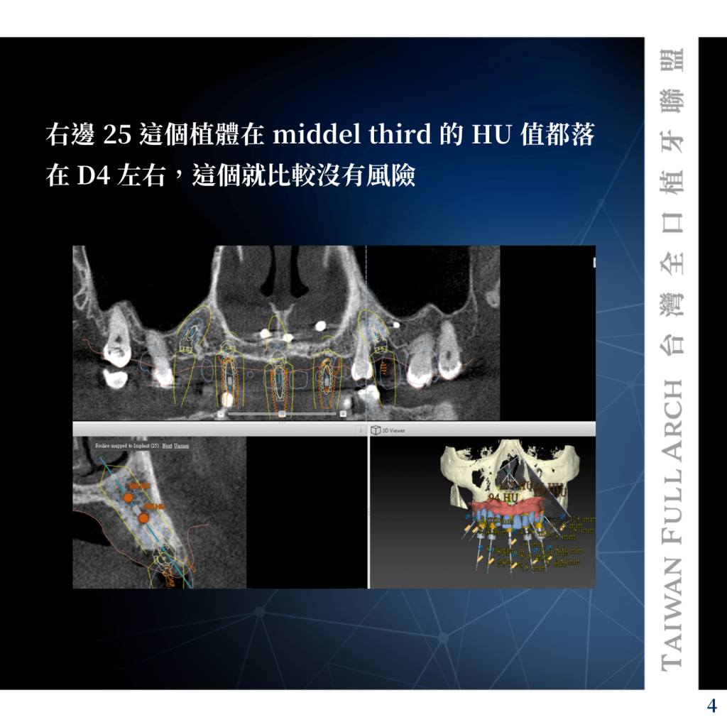
In Figure 2 (right side, implant #25), the HU value in the middle third is approximately D4. This situation carries relatively lower risk.
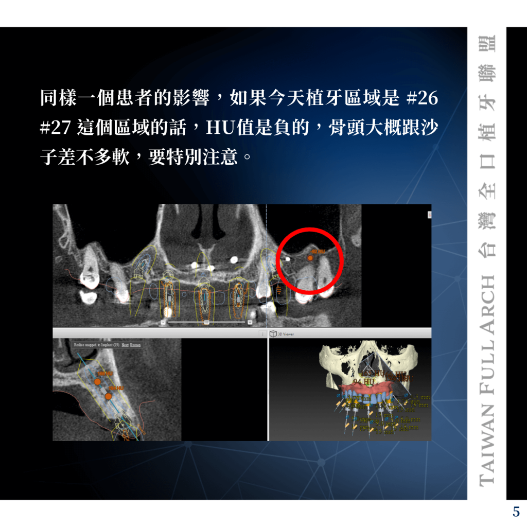
In Figure 3, from the same patient, if the implant site is in the #26–#27 region, the HU value is negative, indicating bone quality comparable to sand. This requires particular caution.
▋ Conclusion
There are now many tools, instruments, and techniques available to manage low-density alveolar bone. However, I believe the most important factor remains accurate imaging interpretation. By mastering this fundamental step and preparing in advance, clinicians can proceed with implant placement more confidently and safely, ultimately fulfilling their commitment to the patient.